


Vatech 3D PaX-i3D
| Optimal FOV Sizes for 3D Diagnosis |
|
|---|---|
| Special Software for each specialty |
|
| Wide Range of Ceph Modes |
|
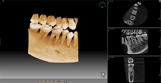
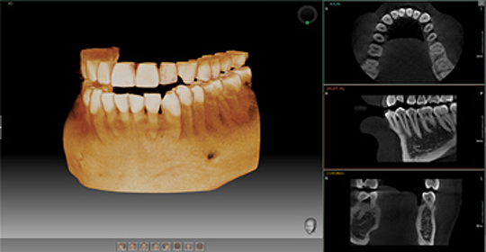
POWERFUL DIAGNOSTIC VALUE WITH 3D IMAGES
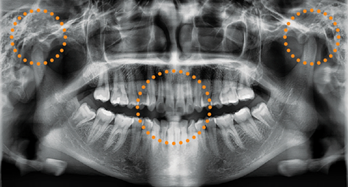
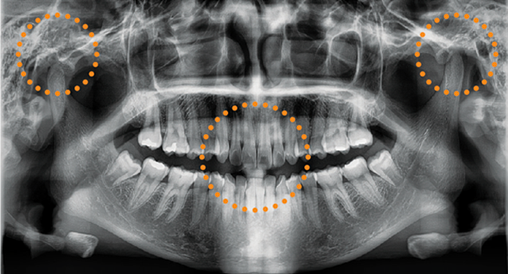
FLEXIBLE 3D IMAGING WITH MULTI FOV SELECTION
PaX-i3D provides 4 multi FOV sizes ranging from 5×5 to 12×9.
By selecting the appropriate FOV size, you can have the optimum image for your diagnostic needs reducing unnecessary X-ray radiation for patients.
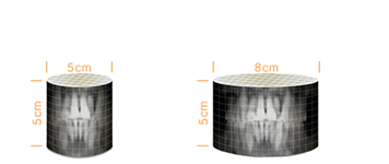
FOV 5X5
5X5 images are useful for specific area diagnosis with minimum X-ray exposure for patients, It can especially increase the accuracy of endodontic diagnosis by exactly checking the amount or root canals and abnormal root canal shapes such as C-shapes that are difficult to check using 2D X-ray system.
FOV 8X5
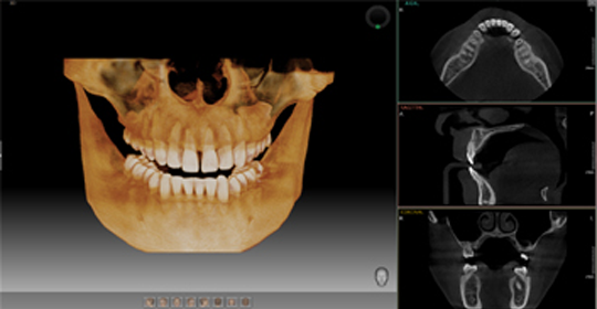
8X5 images can provide more extended oral information on maxillary or mandibular areas. An accurate treatment plan can be established by taking into account the major anatomical structures like mandibular nerve, mental foramen or maxillary sinus.
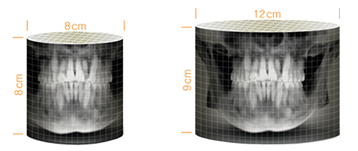
FOV 8X8
8X8 images enable comprehensive diagnosis and treatment planning including both maxillary and mandibular areas in a single scan. It is useful for complex implant surgery as well as left or right TMJ diagnosis.
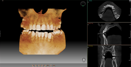
FOV 12X9
12X9 images can provide the most optimal information for oral diagnosis fully covering both maxillary and mandibular structures including the 3rd molar region in a single scan. It is suitable for most oral surgery cases as well as multiple implant surgery.
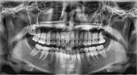
PROFESSIONAL DIAGNOSTIC VALUE WITH PANORAMIC IMAGES
PaX-i3D provides the most precise and high quality panoramic image. Clear and sharp panoramic image brings you better diagnostics. Enhanced details especially in the anterior and dental roots can be viewed. These consistently high quality images will become the new standard of panoramic imaging.
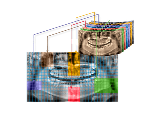
MAGIC PAN
MAGIC PAN creates a more superb panorama image.
It is acquired through the elimination of distorted and blurred images caused by improper patient positioning (Optional).
NORMAL PAN
MAGIC PAN
Focused image is reorganized throughout the whole dental arch and the image quality can be increased.
The image becomes clearer especially in the incisor and canine region, TMJ areas and root canal.
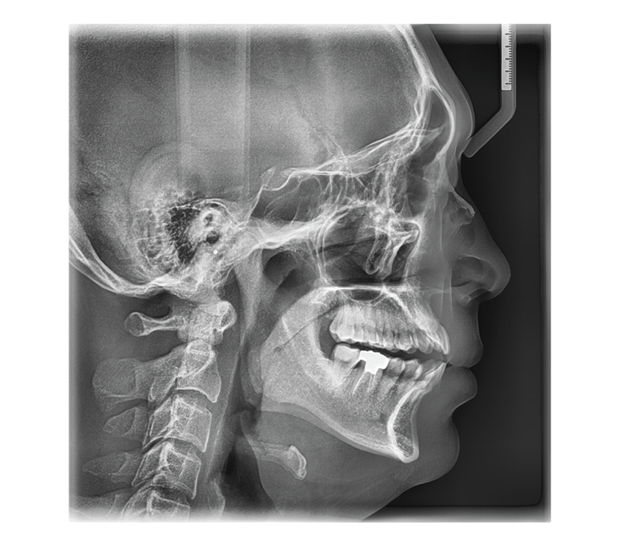
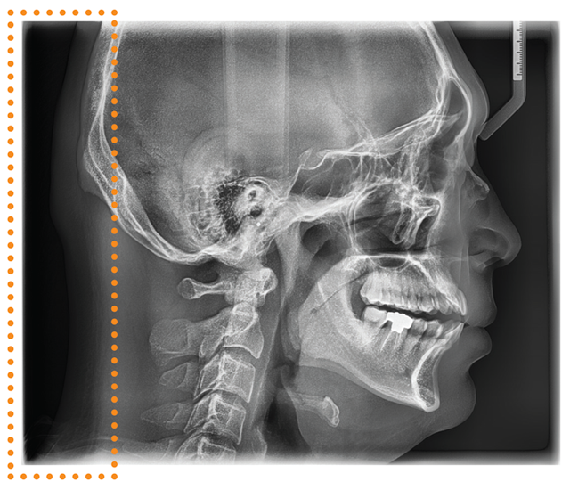
PROFESSIONAL DIAGNOSTIC VALUE WITH CEPHALOMETRIC IMAGES
PaX-i3D Provides optimal images with an exclusively designed sensor for cephalometric diagnosis.
SCAN TYPE CEPHALOMETRIC
Scan type cephalometric offers two image sizes, LAT and FULL LAT, you can choose one of them based on the purposes of your diagnostic needs.
LATERAL
Provide specialized high quality images to suit orthodontics and maxillofacial surgeries.
FULL LATERAL
Full lateral gives 30% larger images and the occipital area of the patient for comprehensive diagnosis. (optional)

-

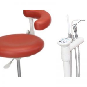
A-dec 300 – Chair-mounted Assistant’s Instrumentation
Delivery Systems, DentalChallenge: Your vacuum delivery solution should comfortably accommodate you and your assistant. Solution: A-dec 300 assistant's instrumentation. The A-dec 300 assistant's instrumentation offers comfortable instrument placement for 2- and 4-handed dentistry. Plus, vacuum instrumentation and other devices are accessible without interfering with instrument transfer or visual or physical oral cavity access. Download Brochure -
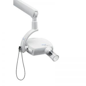
Acteon X-MIND Unity Xray Generator
Acteon, Dental, Feature Product, Imaging, Intra Oral XraysX-MIND Unity - Top Mount (with 80 cm arm)- Smallest focal point on the market - 0.4mm produces a sharp and constrasted image
- ACE Technology reduces radiation exposure by up to 52%
- Clear and large LCD Timer Screen
- Traceability - does received by the patient after each exposure appears on the timer's screen
- With SOPIX inside sensor, this does can also be recorded in the patient's file of Sopro Imaging
- Anti-vibration and anti-movement mechanism ensures drift-free positioning during an exposure
- Selection of the exam type - occlusal or inter-proximal
-
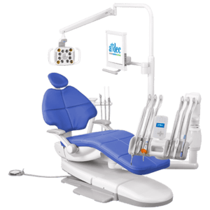
A-dec 400 Dental Chair
A-dec Dental Chairs & Stools, A-dec Showroom Specials, DentalA-dec 400 Dental Chair Instead of sacrificing ergonomic access to the oral cavity for patient comfort, get both with the A-dec 400 dental chair. And when you add robust construction, contemporary styling, and left/right versatility, you gain a system that is the best long-term value available on the market. A-dec 400, a thoughtful approach to dental system design. Take a look at the A-dec 400 Product Brochure -
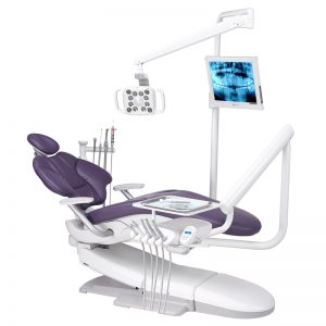
A-dec 500 Dental Chair
A-dec Chair Promo, A-dec Dental Chairs & Stools, A-dec Showroom Specials, Dental, Feature Product
A-dec 500 Dental Chair
A-dec Chair Promo, A-dec Dental Chairs & Stools, A-dec Showroom Specials, Dental, Feature ProductA-dec 500 – a legend, redefined In a world of complex technology and noise, we bring you intelligent simplicity and infallible peace of mind. Designed to function holistically and intuitively, the New A-dec 500 quietly slips into the background, reacting to your every move, without pause. Experience the next level A-dec 500. Take a look at the A-dec 500 Product Brochure [sf_modal header="REQUEST A QUOTE FOR THIS PRODUCT" link_type="button" link_text="Modal link" btn_colour="transparent-dark" btn_type="standard" btn_size="standard" btn_icon="ss-star" btn_text="Request a quote for this product"] [gravityform id="4" title="false" description="false"] [/sf_modal] https://www.youtube.com/watch?v=Cg4UstPwvG8