THE NEW DIGITAL ENVIRONMENT
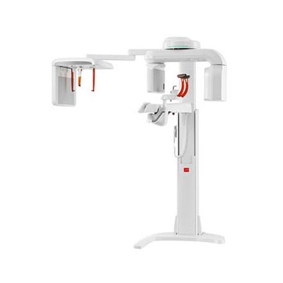
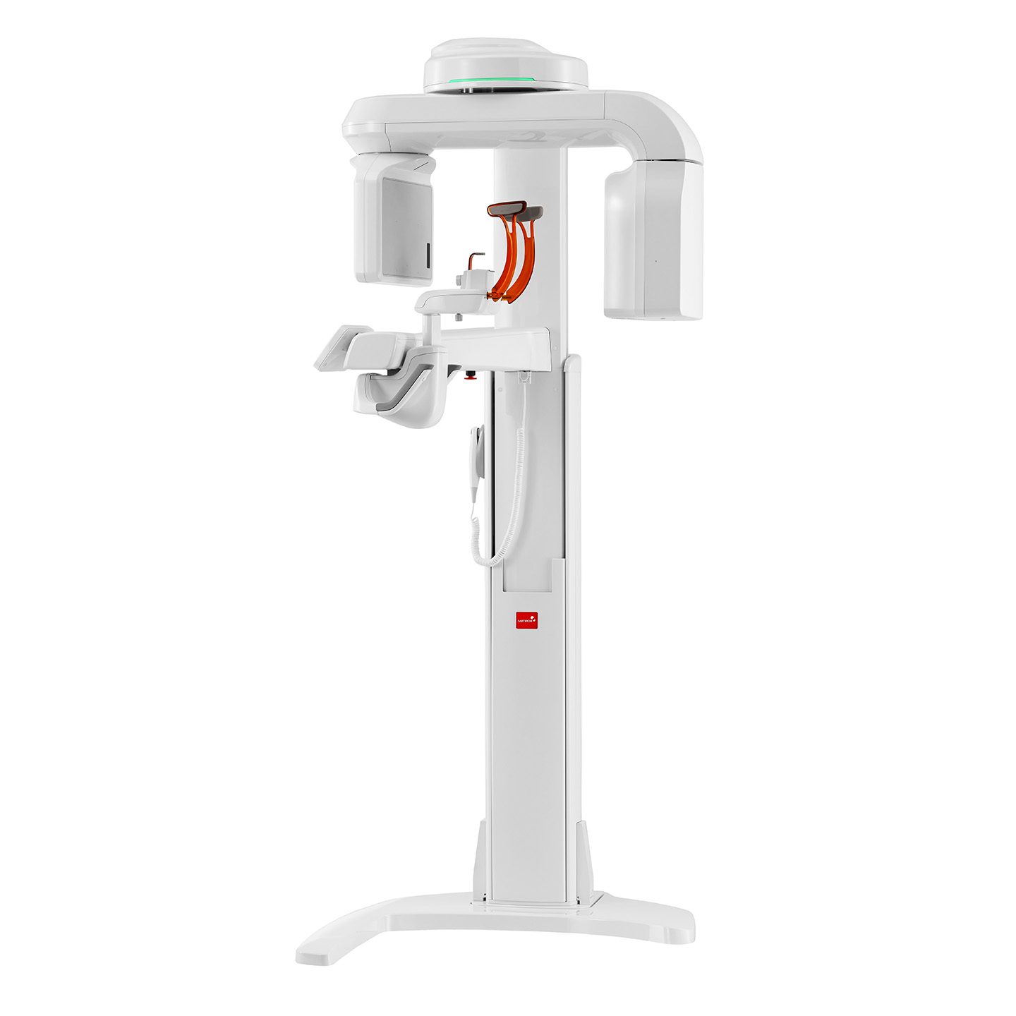
| Green CBCT |
|
|---|---|
| Rapid Scan |
|
| Multi FOV Sizes |
|
| Easy and simple software, Ez3D-i |
|
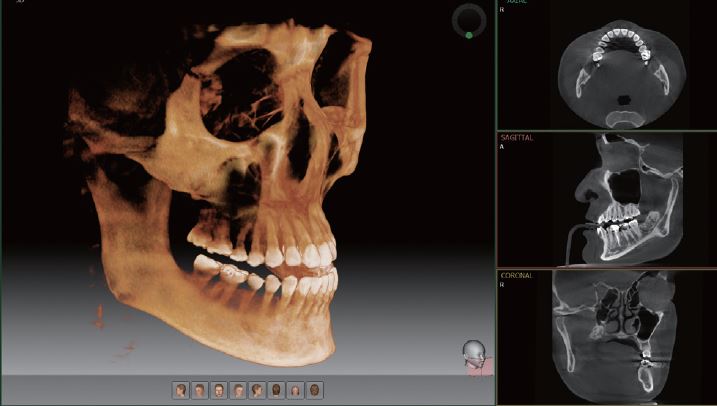
PROFESSIONAL DIAGNOSTIC VALUE WITH 3D IMAGES
Wide range of diagnosis with multi fov selection
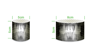
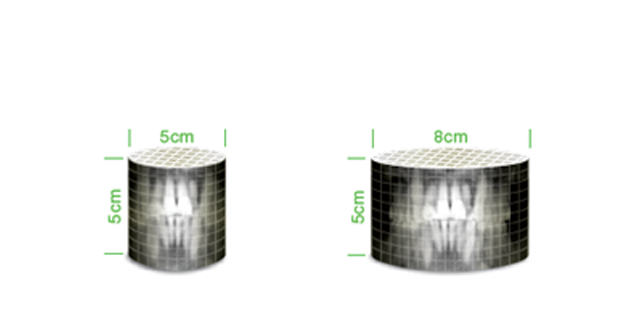
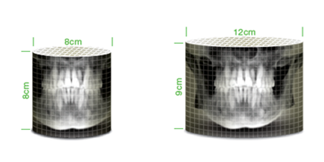
With expanded FOV sizes, the PaX-i3D Green offers valuable diagnoses for professionals.
FOV 10×8
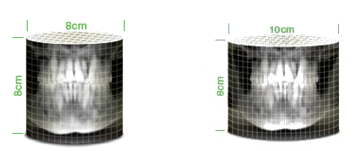
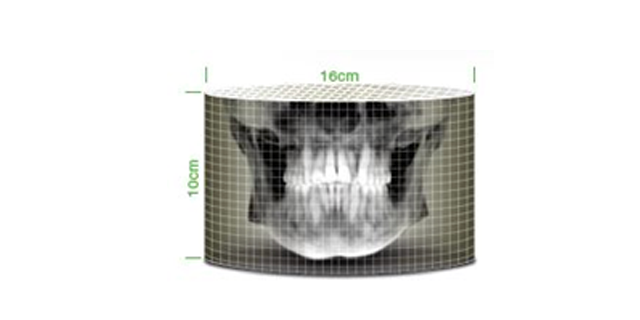
FOV 16×10
FOV 15×15
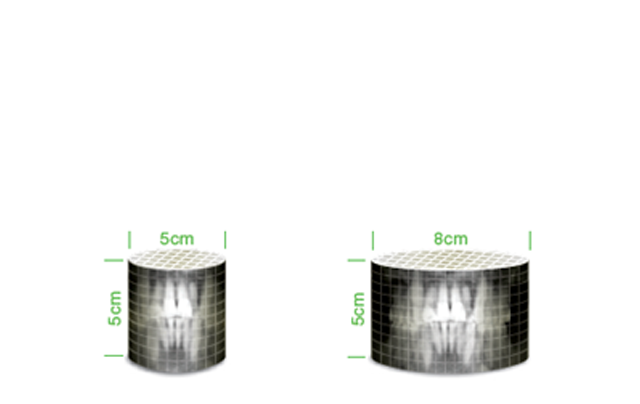
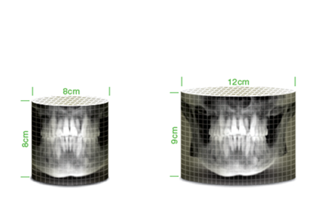
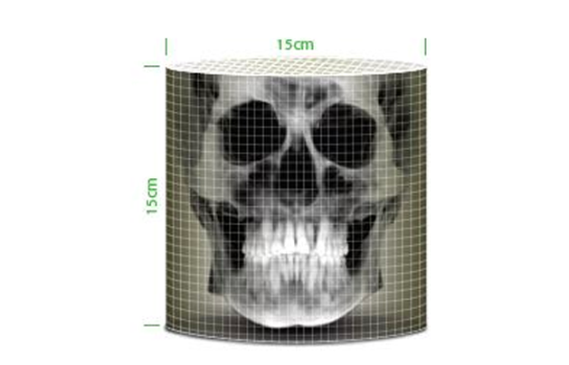
FOV 10X8
10 x 8cm FOV is ideal for dual arch scans of the entire dentition; including third molars, implants and surgical guides.
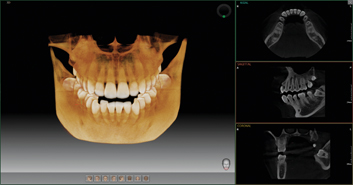
FOV 16X10
16×10 cm FOV provides the optimal information for diagnosis of the entire dental arch, including the TMJ. In addition, implant planning and facial surgery treatment planning is possible.
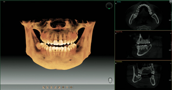
FOV 15X15
15×15 images from PaX-i3D Green enable you to do a comprehensive diagnosis including oral and maxillofacial surgery.
This perfect FOV size will be helpful for complex orthognathic, implant, and orthodontic surgery.
PROFESSIONAL DIAGNOSTIC VALUE WITH PANORAMIC IMAGES
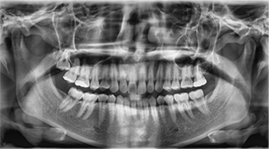
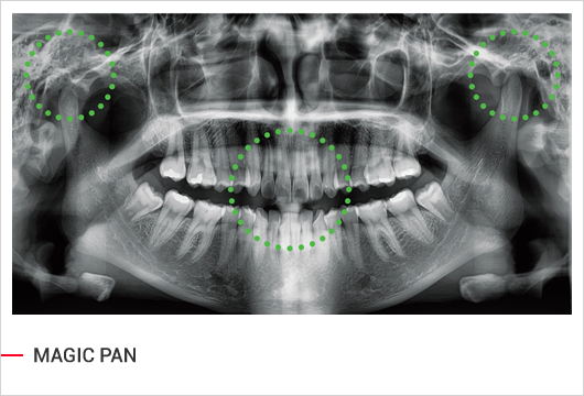
PaX-i3D Green Provides the most precise and high quality panoramic image. Clear and sharp panoramic image brings you better diagnostics. Enhanced details especially in the anterior and dental roots can be viewed. These consistently high quality images will become the new standard of panoramic imaging.
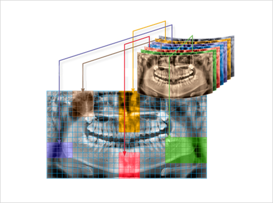
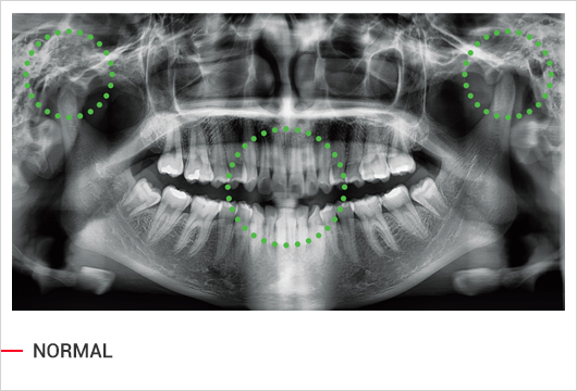
MAGIC PAN
MAGIC PAN creates a more superb panorama image. It is acquired through the elimination of distorted and blurred images caused by improper patient positioning (Optional).
PROFESSIONAL DIAGNOSTIC VALUE WITH CEPHALOMETRIC IMAGES
Extended diagnostic value for wide insight
SCAN TYPE CEPHALOMETRIC
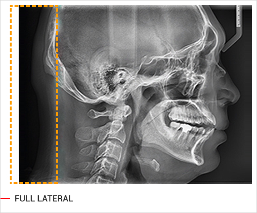
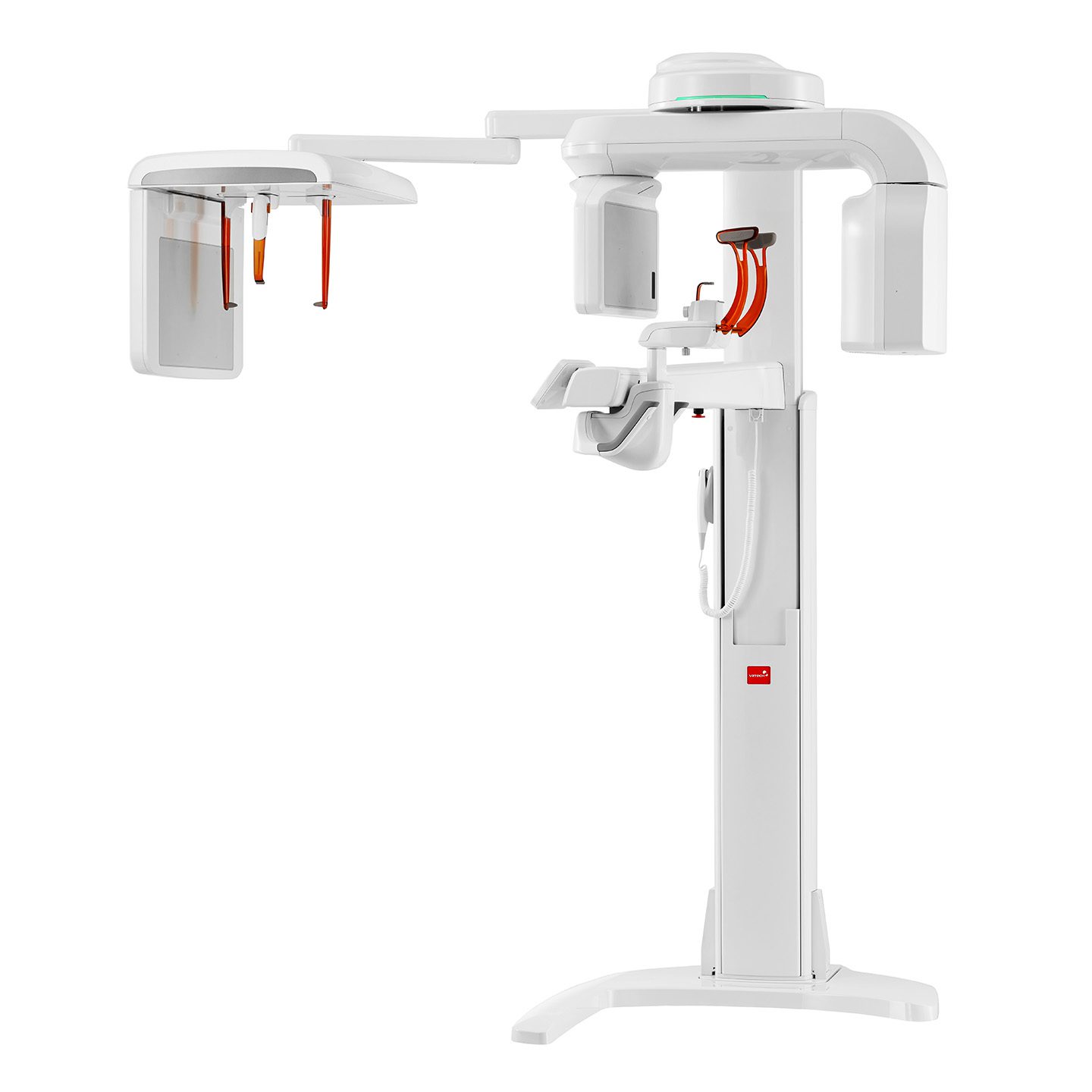
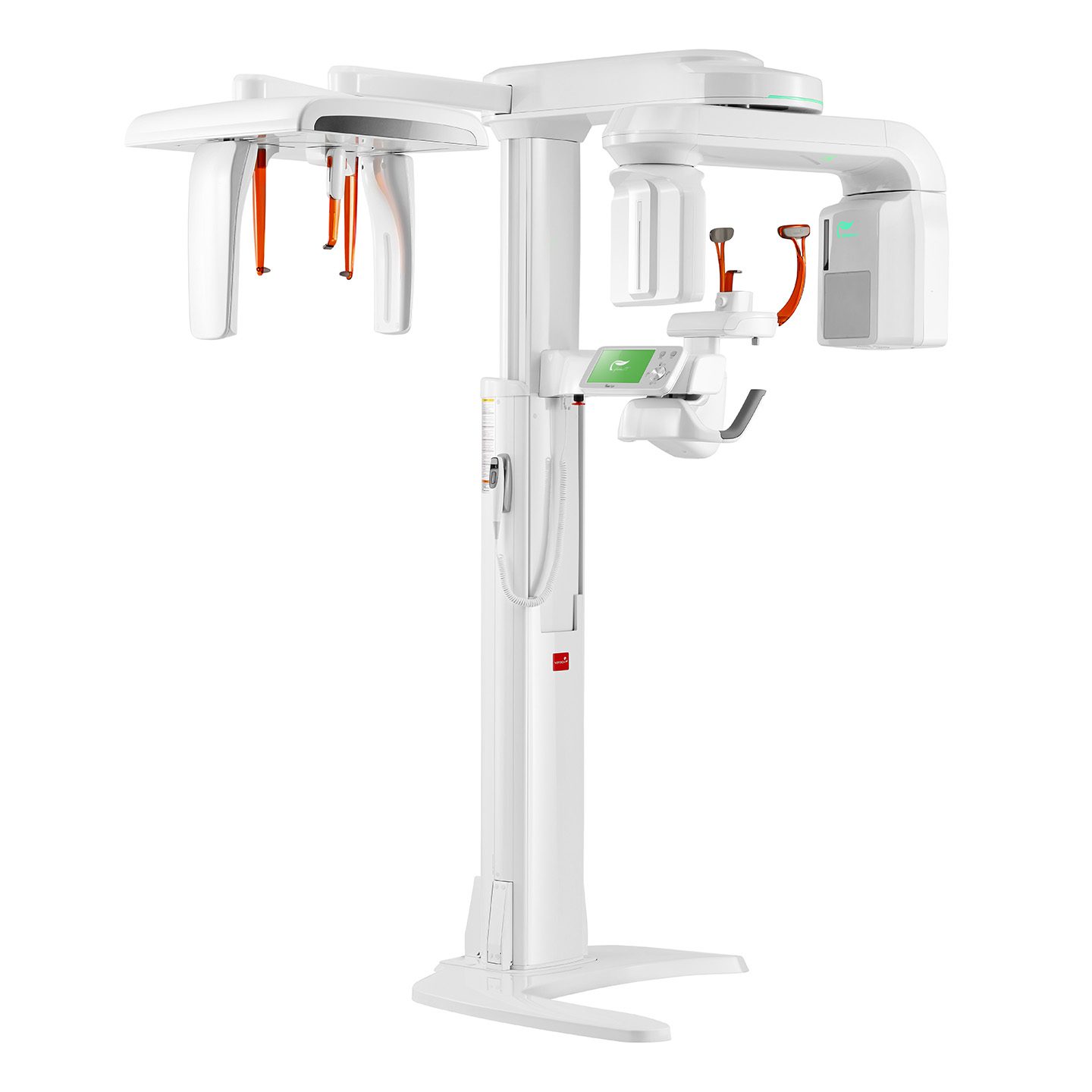
PaX-i3D Green provides optimal images with an exclusively designed sensor for cephalometric diagnosis. As it offers two image sizes, LAT and Full LAT, you can choose one of them based on the purposes of your diagnostic needs.
Built-in Sensor System
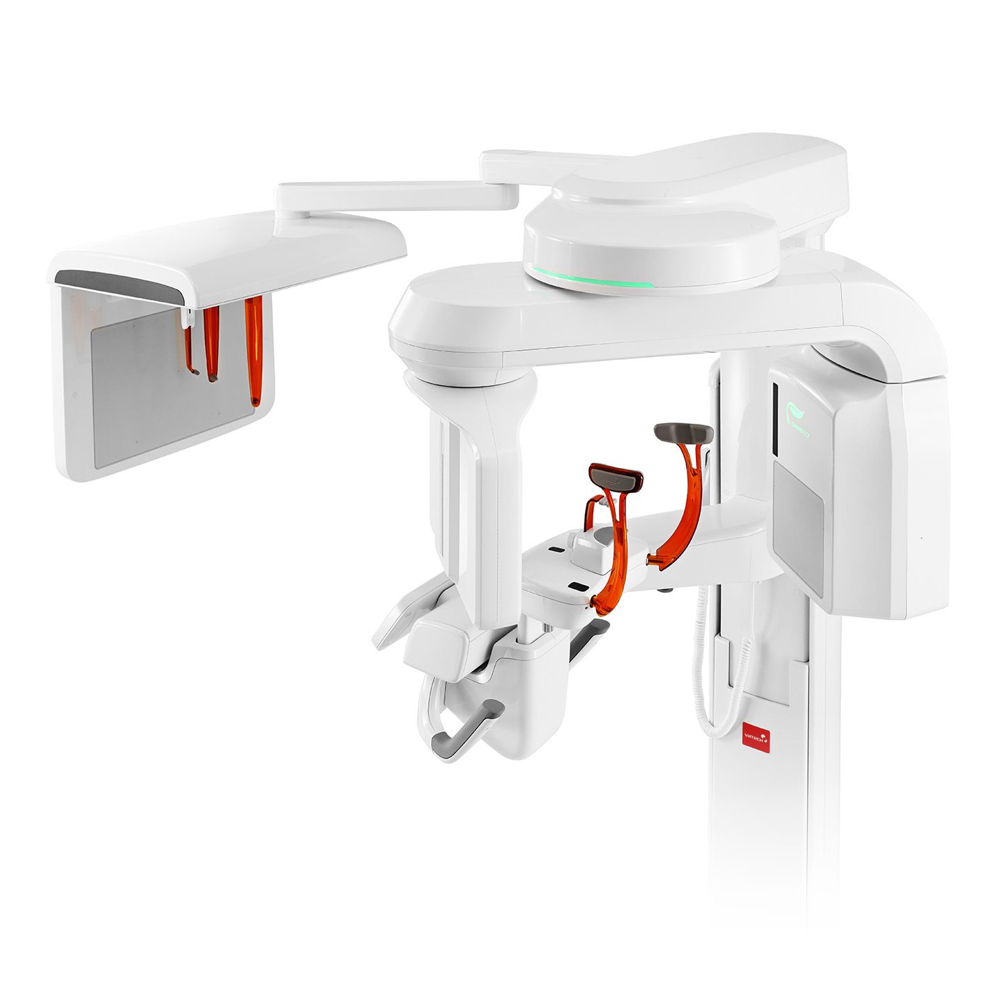
PaX-i3D Green enables you to acquire high quality images in a safe and comfortable environment. Best of all, you don’t need to waste time or risk damage by changing sensors.
Provide specialized high quality images to suit orthodontics and maxillofacial surgeries.
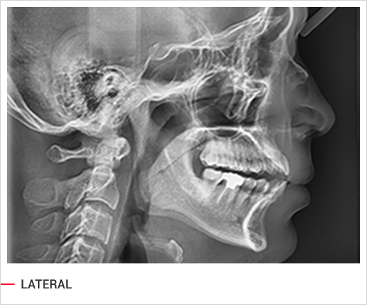
Full lateral gives 30% larger images and the occipital area of the patient for comprehensive diagnosis.
