Durr VistaPano S
VistaPano S – truly perfect imaging
The perfect combination of image quality, efficiency and design
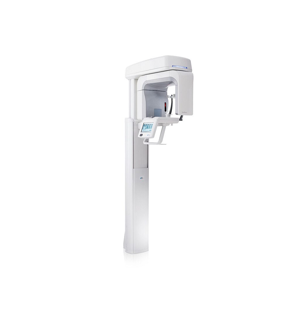
With the VistaPano S, DÜRR DENTAL has set new standards in the resolution of extraoral images. The 2-D panorama X-ray device is impressive with its simple handling and an integrated workflow, supported by an innovative 7″ touch display. The excellent imaging of the VistaPano S is produced by two innovative technologies. Firstly, state-of-the-art Csl sensor technology, producing an improved picture quality, thereby facilitating significantly improved diagnostics. Secondly, the S-Pan technology which uses the patient image to present an automatic razor-sharp panorama of every tooth and jaw position in every situation.
Download Brochure
All functions at a glance
The intuitive 7″ touch display visualises all settings quickly and in high resolution. Handling and navigation are exceptionally user-friendly, ensuring smooth processes while taking X-rays. Simply select the X-ray program and the patient size to prevent handling errors, thereby ensuring an optimal workflow.


Fits in every dental practice
The streamlined, delicate design of the new VistaPano S means it can be placed almost anywhere in your practice. Its compact design with dimensions of 1.0 x 2.3 x 1.5 m (W x H x D) requires very little space and makes it an attractive eye-catcher in any location.


Panorama X-ray programs
With a total of 17 X-ray programs, you are well equipped for every diagnostic requirement. In addition to the standard Panorama program, the VistaPano S offers:
- Half-side recordings of right, left and front
- 4 child programs: an acquisition mode with a smaller exposure area and a 45–56% reduction in the dose without any loss of diagnostic information
- 5 programs for orthogonal X-ray images
- 2 programs for temporomandibular imaging (functional diagnosis)
- 2 programs for sinus X-ray images to display paranasal sinuses


S-Pan technology: extremely sharp images for even more reliable diagnostics
With S-Pan technology, the image regions that best correspond to the actual patient anatomy are automatically selected from a large number of parallel layers. The result is an image of impressive clarity, in which you will be able to immediately and effortlessly locate all anatomically relevant structures. Since the reconstruction is aligned to the actual position of the bite, incorrect positioning is compensated for to a certain extent. This saves time for the surgery and prevents the patient from having to have repeat images taken.



Reviews(0)
There are no reviews yet.